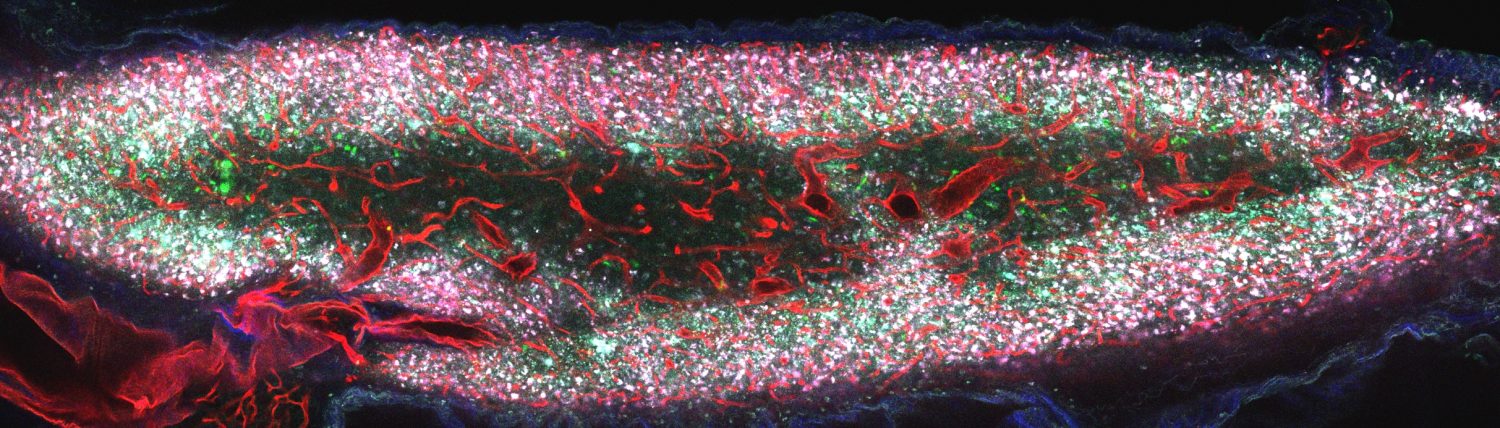
Two-photon RGB image of a mouse thymus after transplantation and tissue clearing
(Red = blood vessels, Green = progenitor cells)
Current Research:
The current research interests of the Spencer Lab for Advanced Microscopy are evenly balanced between the development of novel optical technologies (e.g., two-photon oxygen sensing microscopy) and the application of said technologies to study the dynamic and spatially-varying microenvironmental and cellular factors that influence key biological processes in tissue regeneration and transplantation.
The Spencer Lab’s custom-built two-photon video-rate laser-scanning microscope is designed for live animal imaging. With it, we aim to develop novel intravital imaging techniques for the thymus and to combine them with ex vivo techniques (e.g. cleared whole organ imaging, flow cytometry, single cell RNAseq, etc) to characterize the spatio-temporal changes to the endothelial compartment and surrounding microenvironment (e.g. pO2, blood flow, permeability, etc) after cytotoxic treatment in a level of detail heretofore impossible to obtain with current methods. We seek to identify key microenvironmental factors and novel transcripts important for thymus regeneration.
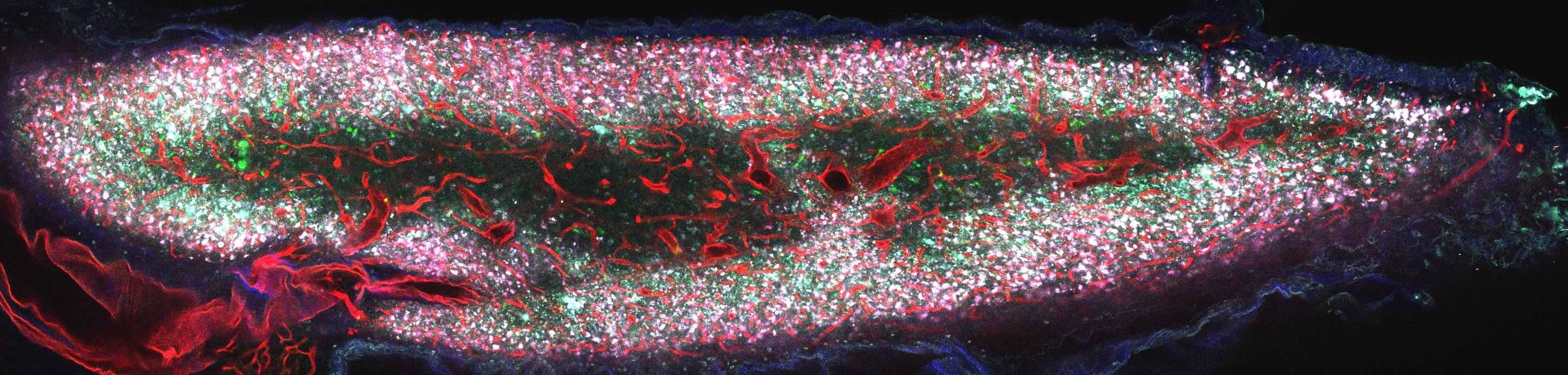
 Two-photon grayscale image of blood vessels in a mouse thymus after tissue clearing
Collaborations
Since these techniques are translatable to other biological contexts, they serve as a starting point for collaboration. We collaborate on multiple diverse projects in hematopoietic stem cell biology, immunology, and beyond.
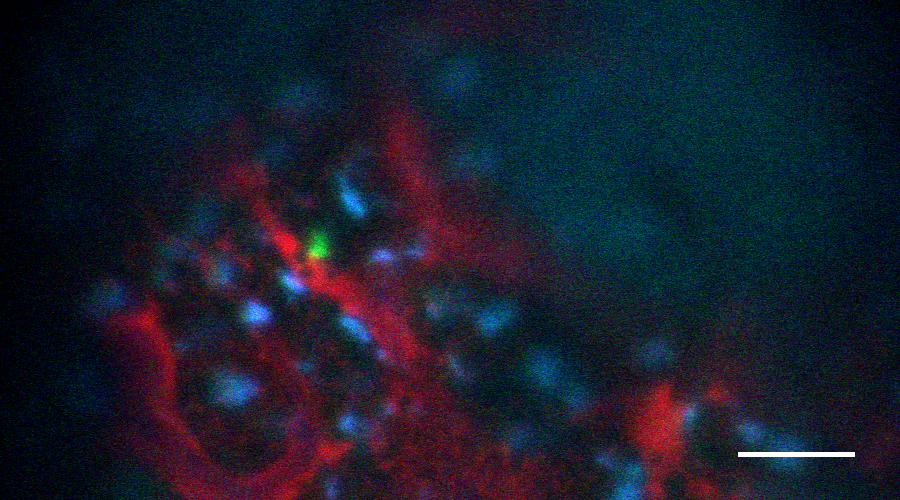
Intravital image of mouse bone marrow (Red = blood vessels, Green = hematopoietic stem cell, Blue = autofluorescence; Scale bar ~50 µm)
Related Videos:
Z-stack movie of the blood vessel network of the whole thymus after tissue clearing:
Stitch_A01_G001_crop_RedchminusBluech_adj_15fps_downsize_con
Time-lapse movie of cell movement within the mouse bone marrow:
TimeLapse5minStack_0to4i_adj_timestamp_50umSB_adj (Converted)